
South African doctor at the centre of cutting-edge technology transforming fetal surgery in US
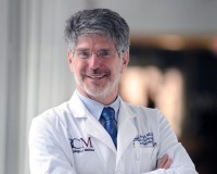
Dr Michael A. Belfort, MD, PhD, Chair of the Department of Obstetrics and Gynecology at Baylor College of Medicine (BCM) and Obstetrician and Gynecologist-in-Chief of Texas Children's Hospital.
Dr Michael Belfort, Obstetrician and Gynaecologist-in-Chief at Texas Children's Hospital and a specialist in maternal-fetal medicine and fetal intervention, led a team that performed minimally invasive surgery on an unborn baby diagnosed with complex gastroschisis. The procedure, performed when the baby was 26 weeks into gestation, uses technology more commonly associated with keyhole surgery but adapted for an extremely challenging environment: the inside of the womb. Taking keyhole surgery into the womb Gastroschisis occurs when a baby's intestines develop outside the abdominal wall. In conventional treatment, the exposed intestines are protected after birth and surgeons subsequently return them to the abdomen. The Texas team is investigating whether advanced fetal intervention can address the problem before delivery. The procedure uses fetoscopic surgery, a minimally invasive technique in which surgeons access the uterus through tiny ports rather than making a large opening in the uterus. The technology allows a miniature camera and specialised surgical instruments to enter the womb. Surgeons can then view and manipulate the fetus while relying on highly controlled surgical access and imaging. Texas Children's has extensive experience with this type of technology. Its fetal surgery programme has previously developed a two-port fetoscopic technique for repairing spina bifida, using tiny uterine openings, a fetoscope and miniature instruments. Carbon dioxide replaces amniotic fluid One of the most striking technological elements of the procedure is how surgeons create a workable environment inside the uterus. During the fetoscopic procedure, some of the amniotic fluid is removed and replaced with carbon dioxide gas. This creates a clearer operating environment, allowing the surgical team to see and manipulate structures inside the womb. The approach is based on technology used in minimally invasive surgery, but it has been significantly adapted for fetal intervention. The surgical instruments themselves are extremely small, reflecting the scale at which fetal surgeons must operate. This is not simply conventional surgery performed on a smaller patient. The technology has to account for the movement of the fetus, the mother's anatomy, the uterus, amniotic environment and the need to protect both mother and baby. A decade of technological development The procedure is the result of years of research and technological development. Belfort's experience with fetoscopic treatment for spina bifida helped establish a platform for exploring new applications of fetal surgery. Texas Children's says its team has performed more than 220 fetoscopic spina bifida repairs as of November 2025. The development process has also involved simulation and animal research before moving into human clinical trials. Texas Children's has described how its fetal surgery team used simulation models to refine the technique before progressing to sheep models and eventually human procedures. That same philosophy is now being applied to complex gastroschisis. The current gastroschisis programme is being studied through a formal clinical trial designed to assess the safety and feasibility of fetal repair using a fetoscopic surgical approach. Technology could change what happens after birth The potential impact goes beyond the operation itself. Severe or complex gastroschisis can cause significant intestinal damage. Babies may require prolonged neonatal intensive care, multiple procedures and extended nutritional support after birth. The theory behind fetal intervention is that repairing the condition earlier could reduce damage to the exposed bowel and potentially improve the baby's condition by the time of delivery. Texas Children's currently uses advanced prenatal imaging, including detailed ultrasound and fetal MRI, when assessing babies with gastroschisis. These technologies help specialists understand the severity of the condition and plan treatment. The emerging combination of advanced imaging, fetoscopic cameras, miniature surgical instruments, specialised access systems and minimally invasive surgical techniques represents a new generation of fetal medicine. Could South Africa adopt the technology? For South Africa, the development raises an important question: could this technology eventually be used locally? Belfort, who was born in South Africa and trained at the University of the Witwatersrand and University of Cape Town, believes the country has much of the expertise and infrastructure required to develop an advanced fetal surgery programme. However, establishing such a service would require more than purchasing specialised equipment. It would require a highly coordinated multidisciplinary team involving maternal-fetal medicine specialists, fetal surgeons, paediatric surgeons, neonatologists, anaesthetists, imaging specialists, nurses and other technical professionals. Texas Children's own model demonstrates the importance of bringing these disciplines together in a single specialised environment. There is also a significant research and regulatory component. Fetal surgery involves two patients — the mother and the fetus — meaning new procedures must be carefully evaluated for both maternal and fetal safety. The future of fetal medicine The gastroschisis procedure illustrates how medical technology is increasingly moving treatment from the neonatal intensive care unit into the womb itself. What once required surgeons to wait until birth can increasingly be approached through sophisticated fetal imaging, minimally invasive instruments and specialised surgical platforms. For Belfort and his colleagues, the goal is not simply to demonstrate that the procedure can be performed. The ongoing clinical trial is intended to establish whether fetal repair can safely improve outcomes for babies with complex gastroschisis. If future evidence confirms its benefits, the technology could eventually become another tool available to specialists treating serious conditions before birth. For South Africa, the story also highlights an opportunity: the country already has world-class medical expertise in several areas, and with the right investment, infrastructure and multidisciplinary collaboration, advanced fetal intervention could become part of the country's future healthcare technology landscape.